The Importance of Good Positioning on Canine Hip X-rays
The purpose of this article is to teach the average dog owner how to determine if a hip x-ray is done properly on their dog's hips. The article will demonstrate correct positioning and poor positioning. It will show 2 different sets of x-rays done on the same dog on the same day. One set has good positioning, the second set has poor positioning. You will see that with poor positioning, a dog's hips can look worse than they actually are. You will also see that no matter what you do with positioning you can never make a bad hip into a good hip.
Over the years, I have seen some absolutely terrible jobs of x-raying dogs. As time goes by, I will continue to add examples of poor x-rays to this article so people can learn what to look for.
How to Read a Hip X-Ray
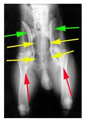
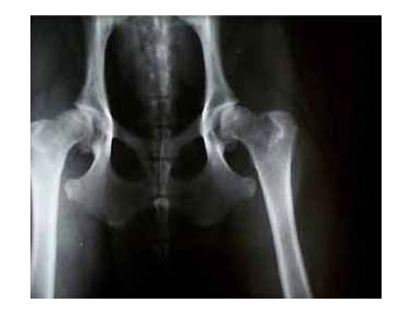
Figure 1 is a photo of a hip x-ray done on a 10-month-old German Shepherd from my kennel. While the dog is slightly angled on the x-ray plate, the positioning for the hips is pretty good. It shows the various points on an x-ray to look at to determine if the dog was positioned properly.
Because this article is directed to the general public, I will attempt to not use the proper medical names for a lot of the terminology in this article.
The first thing to look at in an x-ray is to see if the legs come straight down from the hips with the kneecaps square and looking alike. We don't want to see one leg straight and the other going off at an angle.
Figure 1 has three sets of colored arrows (green, yellow, and red).
The green arrows point to the bone that the hip socket is built into. These bones almost look like wings. You will notice that you can see more of the wing on the right than the wing on the left. When the position is 100% perfect, both wings will look exactly alike.
The yellow arrows point to holes in the bone structure. When the body positioning is correct, the 2 holes on the left side are the same shape and size as the holes on the right side. The positioning is good on this dog, but not 100% perfect. That's why the holes on the right are slightly different than the left. This is most noticeable in the lower right hole being smaller than the left side lower hole.
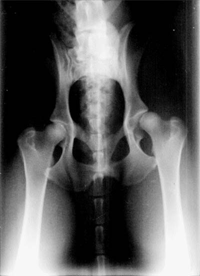
The red arrows are the first things I look at when examining an x-ray. They point to the amount of pelvis bone that is covered by the leg bones on the x-ray. If you look at the pelvis, you can see that with the legs fully extended straight down, the legs overlay the very corners or tips of the pelvis. You can see the overlap through the leg bone. The picture above shows an even amount of overlap on both sides of the pelvis. Figure 2 shows a much larger overlap on the left of the screen than on the right of the screen. This is poor positioning.
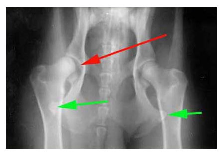
Figure 2 above is the same dog only with a different x-ray than the first one. This second x-ray has poor positioning. Notice how much more the pelvic overlaps the leg bone (the green arrows) on the left than on the right. The result is the hip is pulled further out of the socket (the single red arrow) because of poor positioning.
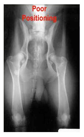
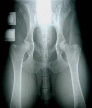
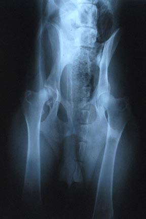
Figure 3 is an example of poor positioning. Again this is the same dog as the good x-rays above. The dog is rotated. You can see the upper right hole through the body cavity is noticeably smaller on the right than the left. The pelvic wing under the leg is noticeably larger on the left than the right.
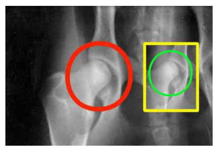
Figure 4 graphically shows the results of poor positioning. It shows the same hip joint on the same dog x-rayed on the same day. The hip in the red circle is a much deeper seated ball in the socket than the picture in the yellow box (which is a result of poor positioning).
Some people ask how the difference can be so dramatic. My feeling is that these are young dogs. They have loose ligaments (just like a young child). If I took some of the falls that my eleven-year-old son does, I would have numerous broken bones. It's the same with our dogs. As they get older, their ligaments are not as loose and they will probably not stretch as much. There may not be as much of a difference in older dogs. But at a young age, positioning is critical.
The importance of positioning is often overlooked by the vet that is shooting the films. There may be a number of reasons for this:
- It could be a lack of experience doing hip x-rays.
- It could be a money issue with him. Shooting another x-ray because he made a mistake costs him money.
- It could be that by the time the x-ray is developed and he realizes the position wasn't that good, the animal is gone or awake from being knocked out.
In my opinion, none of these are good reasons. To get good x-rays, you have to have a good vet. I have a couple of local vets that are very good with x-rays. If they make a mistake, they re-shoot it at their expense. We just recently started to see the OFA send x-rays back to the vets because of poor positioning. When this starts to happen on a consistent basis, we will start to see much better x-rays of the dogs.
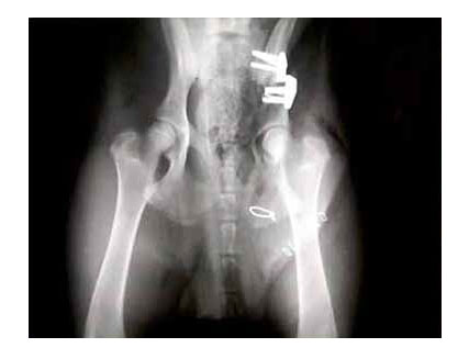
There are several operations that are being done today to correct a bad hip and allow the dog to live a normal life. Figure 5 is an example of what a hip can look like after the operation. This operation needs to be done at an early age.
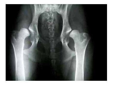
Figure 6 shows a very bad set of hips. It's questionable if surgery could even correct this dog's problem. These are hips from an 8-month old German Shepherd that came from a backyard breeder. A dog with hips like this should be put down. It is facing a life of pain.
Figures 7 and 8 above are of the same dog, a Border Collie. Figure 7 was taken at 8 months of age. Figure 8 was taken at 4 years of age. This can give you an idea of what will happen to bad hips over time. Notice the thickening of the neck of the joint. The ball also shows signs of arthritis. This dog is living as a house dog where her exercise is monitored. When the pain gets bad, she is given Rymadil and this seems to make her comfortable.
My advice to anyone would be to not accept incorrect positioning of any kind. Discuss this with the vet before the x-ray. Show him this article if he has any questions. I personally will not pay for a bad x-ray.
I recently had a similar situation with a young dog that I x-rayed at 6 months. The picture did not look that good but the rest of the litter was good. So I redid the x-ray at 9 months and saw an entirely different x-ray. The dog will pass OFA if the x-ray stays the same.
I would also recommend swimming a dog to build muscle mass if there is any question on the hips. The better condition a dog is in, the better chance of a good x-ray. I have a friend who has watched the OFA on a yearly basis. She has noticed that there are more bad hips in the winter months than summer months.
For me, this translates into dogs not being in as good physical condition in the winter months as in the summer. In the future, I will not be x-raying dogs in the winter. I will also make sure that my dogs are in excellent condition when the x-rays are taken.
Examples of Hip X-Rays
Example 1
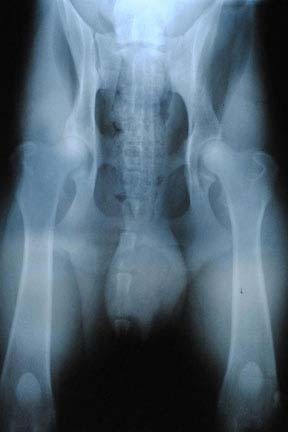
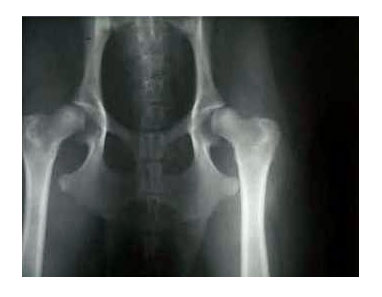
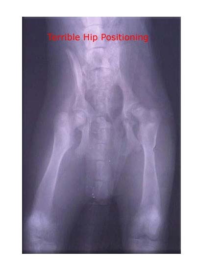
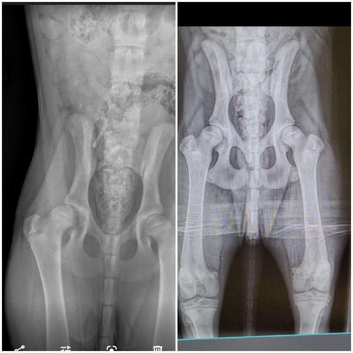
Figures 11 and 12 are x-rays of the same dog. Figure 11 displays poor hip positioning. Figure 12 has the correct hip positioning. Both x-rays were taken just several days apart.
While this dog does not have perfect hips, if you look at the positioning of the x-ray on the left, the hip positioning makes the dog's hips look much worse than they really are. This was confirmed when the dog's owner took this article back to her vet and asked the vet to re-do the x-ray with correct positioning.
Example 2
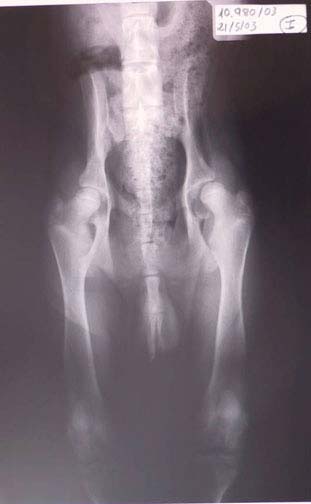
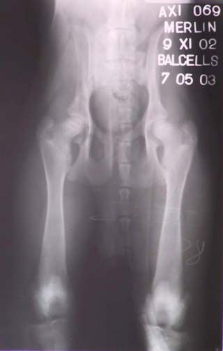
Figures 13, 14, and 15 are x-rays of the same dog done at different times. Positioning is important when it comes to taking x-rays. Look at the right hip in all three shots. You will notice that Figure 13 is much different from Figure 15.
Example 3
Figures 16 is an image of x-rays sent to me by a customer in January 2022. They wrote, "I read a story on one of your forums and you talked about good positioning. The x-rays are from a Dutch Shepherd pup I sold. The left is from the new owner's vet and the right is from my vet while the pup was sedated. Just thought it was worth a mention, because of you, I decided to get a second opinion."
The Worst Positioned Hips I Have Seen
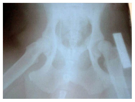
Figure 18 was sent to me in February 2006. They are the worst example of hip positioning I have ever seen. The vet that took these should give up his day job and seek another career.
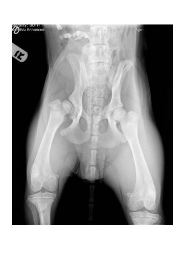
Figure 19 shows awful hip positioning by the vet. The hips are bad, however. No matter how they were positioned, it would not have made them look any better.
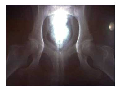
Figure 20 is one of the worst case of hip positioning that I have ever seen. The vet that took them and gave them to the customer should get out of the business.
How to Prevent Bad Hips in Dogs
With all this said, if you are reading, you are probably asking yourself what you can do to make sure your dog has healthy hips. The SV in Germany (the German Shepherd Dog Club of Germany) has proven that genetics is only responsible for about 25% of the bad hips in dogs. This means that 75% of bad hips are caused by environmental issues.
There are things that help:
- Keep your dog thin. When I say thin, I mean you need to see a definition between the ribs and loins of your dog. I cannot stress this enough. The more weight a dog carries, the more pressure on the hips. This is extremely important when the dog is growing (between 8 weeks and 18 months).
- Do not over-exercise your young dog. DO NOT TAKE A PUPPY JOGGING! You must wait until it's older than one year of age. Overly exercising your puppy is the fastest way to destroy hips.
- Feed a quality all-natural diet. If you don't want to feed a raw diet, at least feed an all-natural kibble diet. We also sell all-natural food options which you can find here. The brands we carry and sell are what we feed to our dogs. We stress the importance of a proper diet with our puppy customers and it has made a huge difference
- If you have a question about subluxation in a young dog, SWIM the dog! Take the dog swimming every day for 3 or 4 months before you have x-rays taken. Swimming is the best exercise you can do for a dog. It is way better than jogging the dog. A subluxation in a dog means the head of the femur is loose in the socket. It makes sense to exercise the dog so that the muscles and ligaments tighten up the bones as much as possible.
- We give our dogs hip and joint supplements. View some of the products we use.
Unfortunately, you can do all of the things mentioned above and still get bad hips. That's the sad thing. I have bred over 350 litters in 30 years. The dogs I breed have good hips 6 to 10 generations and we will occasionally get a bad hip. However, I will say that the percentage of hip problems in our kennel is much, much less than breeders who do not follow this protocol.
Additional Reading
- Notes from the Tufts Genetics Breeders Conference
- Chronology of Hip Dysplasia Development in a Cohort of 48 Labrador Retrievers Followed for Life


Most Popular Articles
-
01
-
02
-
03
-
04
-
05
Related Products
-
 $65.00
 -
 $16.99
 -
$219.99 - $349.99
$199.99 - $309.99 -

$65.00$19.99

Ask Leerburg.