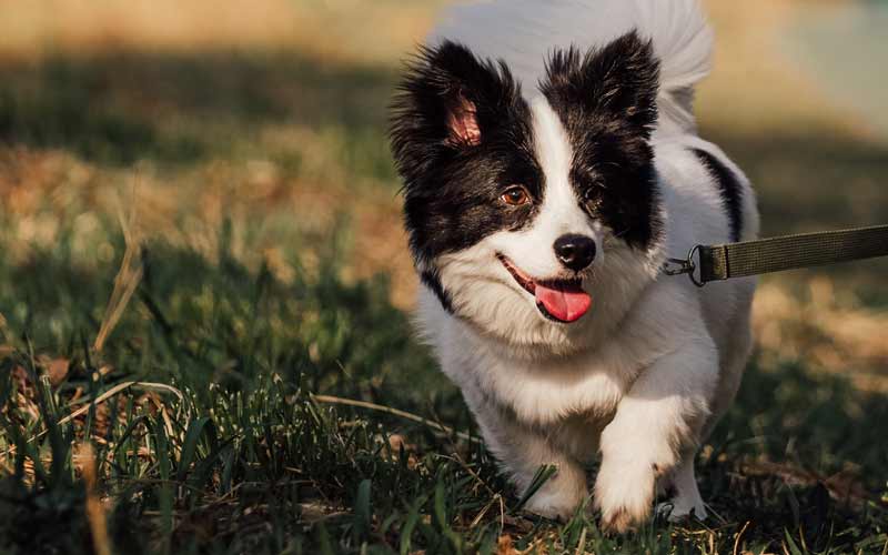
Elbow Dysplasia
![]() |
WHAT IS ELBOW DYSPLASIA?
Elbow dysplasia was originally described as a developmental disease manifested as degenerative joint disease of the elbow with or without an ununited anconeal process. Erroneously, the term elbow dysplasia became synonymous with ununited anconeal process thus causing the confusion that exists among some veterinarians and breeders.
Developmental degenerative joint disease of the elbow has multiple inherited etiologies, which may occur singularly or in combination (ununited anconeal process, fragmented medial coronoid process, or osteochondritis of the medial humeral condyle).
METHOD OF DIAGNOSIS
Proper evaluation of this region requires the elbow be positioned in extreme flexion and good radiographic technique be used. If a specific etiology is sought additional views are suggested.

Extreme flexed lateral required
The purpose of the registry is to identify phenotypically normal dogs and screen elbow radiographs for signs of early degenerative joint disease. The earliest and most consistent secondary change is a smooth periosteal reaction on the proximal anconeal process and/or joint incongruity.

Periosteal reaction on anconeal process
Normal elbows on individuals 24 months or older are assigned a breed registry number and will periodically be reported to the parent breed club.
Abnormal findings are reported only to the owner of record and referring veterinarian.
Abnormal elbows are reported as:
- Grade I- minimal bone change on the anconeal process.
- Grand II- additional subchondral bone changes and/or
osteophytes.
- Grade III- well developed degenerative joint disease.

Craniocaudal

Neutral Lateral

15-20 degree Craniolateral-Caudomedial Oblique
TESTIMONIAL:
While surfing your website, I came across the article on elbow dysplasia. I have had first hand experience with this condition. After talking with the broker where my department purchased the dog, he had never heard condition. Likewise, I was unable to find anyone else in the area K9 programs who have heard or dealt with this condition. I would like, to share my experience with you.
I am a K9 handier for the Rockford (Illinois) Police Department. In November, 1998, 1 graduated with my partner, Valco, a 2 1/2 year old Dutch Shepherd from the Illinois State Police Academy K9 training section. A few times during class, I noticed that Valco was limping. I would check his left leg and noticed no problems. Valco would also show no complaints to my manipulation of his leg. I had him checked by my vet who could likewise find no problem on external exam. Due to the training, the problem was figured to be a muscle strain. Valco was put on Rimadyl and continued to train.
In late December'98, Valco began limping again, but this time on a daily basis. He again showed no complaints upon examination by me or my vet. X-rays were taken in early January and Valco was diagnosed with Ununited Anconeal Process. Very slight arthritis was also evident in the joint. I took Valco to the University of Wisconsin (Madison) Vet Hospital where he was examined by orthopedic surgeons. After much debate with my department, the decision was made to have screw fixation surgery to reattach the anconeal process to stabilize the joint and try to continue working Valco as a full service dog. Valco was returned to the UW for surgery. I was notified that during workups for surgery, x-rays taken at the UW also showed a high probability of Fragmented Medial Corornid Process. The decision was made not to have surgery but replace the dog as he was still under skeletal warranty from the broker.
This is a problem I had never heard of until my dog was diagnosed with it. I am sure most handlers have never heard of it either. It is not a common affliction, but it is a career shortening or ending problem for the dog. Attached is a photo of Valco. If you look closely at his left elbow, it is bowed outward slightly. This photo was taken 6 days before he was diagnosed.
Respectfully,
Mark
Rockford Police Dept.


